microarray banner

Karyotyping - For Doctors

Home / For Doctors / Karyotyping - For Doctors

Karyotyping for Genetic Diagnosis

Karyotyping is a fundamental cytogenetic technique used to visualize and analyze an individual's chromosomes. It allows for the detection of numerical chromosomal abnormalities (aneuploidies) and large-scale structural rearrangements (e.g., translocations, deletions, duplications, inversions).

While newer technologies like chromosomal microarray (CMA) offer higher resolution for detecting copy number variations (CNVs), karyotyping remains a crucial diagnostic tool in various clinical settings, particularly for identifying balanced rearrangements and confirming suspected aneuploidies.
what is microarray

Key Applications

Indications for Karyotyping

Karyotyping is indicated in a range of prenatal, postnatal, and specialized scenarios:

Prenatal Diagnosis (from Amniotic Fluid - AF, or Chorionic Villus Sample - CVS):

  • Positive prenatal screening results (e.g., NIPT, first/second-trimester serum screening) suggesting aneuploidy.
  • Fetal structural anomalies detected on ultrasound.
  • Fetal structural anomalies detected on ultrasound.
  • Previous child with a chromosomal abnormality.
  • Advanced maternal age (though NIPT/CMA are often primary).
  • Family history of a condition diagnosable by karyotyping.

Postnatal Diagnosis (from Peripheral Blood):

  • Suspected aneuploidy syndromes (e.g., Down syndrome, Turner syndrome, Klinefelter syndrome).
  • Individuals with developmental delay (DD), intellectual disability (ID), or autism spectrum disorder (ASD), often as a complement to CMA.
  • Multiple congenital anomalies (MCA).
  • Disorders of sex development (DSD).
  • Primary amenorrhea or infertility (male and female).
  • Short stature of unknown etiology.

Other Applications:

  • Recurrent Pregnancy Loss (RPL): Parental karyotyping to detect balanced translocations or inversions.

Conditions Detected by Karyotyping:

Numerical Abnormalities (Aneuploidies):

Structural Abnormalities (if large enough, typically >5-10 Mb):

Mosaicism: Presence of two or more cell lines with different chromosomal constitutions (detection depends on the percentage of mosaicism and number of cells analyzed).
Platform & Methodology

Methodology: G-Banded Karyotyping

  • Sample Collection: Blood (in Sodium Heparin), AF, CVS.
  • Cell Culture: Cells (lymphocytes, amniocytes, chorionic villi cells, fibroblasts) are cultured in appropriate media to stimulate cell division.
  • Mitotic Arrest: A mitotic inhibitor (e.g., colcemid) is added to arrest cells in metaphase.
  • Hypotonic Treatment: Cells are treated with a hypotonic solution to swell them and disperse chromosomes.
  • Fixation: Cells are fixed, typically with a methanol:acetic acid fixative.
  • Slide Preparation: Fixed cells are dropped onto microscope slides and air-dried.
  • Banding: Slides are treated with trypsin and stained (e.g., Giemsa stain) to produce characteristic G-bands on chromosomes.
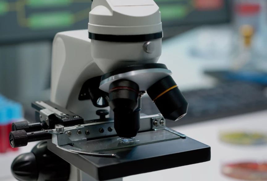
  • Microscopic Analysis: Chromosomes are visualized, counted, and analyzed for structural integrity by a trained cytogeneticist. Images are captured, and a karyogram is constructed.

Precision & Progress

Understanding Karyotype Results

Shipping Conditions : Room temperature or cold pack (depending on sample type).
Normal Female:

46,XX

Normal Male:

46,XX

Numerical Abnormality:

e.g., 47,XX,+21 (Female with Down syndrome)

Structural Abnormality:

e.g., 46,XY,t(9;22)(q34;q11.2) (Male with a balanced translocation between chromosomes 9 and 22)

Mosaicism:

e.g., mos 45,X[10]/46,XX[10] (Mosaic Turner syndrome with 10 cells showing 45,X and 10 cells showing 46,XX))

Microarray recommended

Clinical Guidelines

Clinical Guidelines Supporting Karyotyping Use

  • ACMG/ACOG: Recommend karyotyping for specific prenatal indications (e.g., confirming NIPT, parental translocation) and in certain postnatal evaluations (e.g., suspected aneuploidy, RPL workup).
  • Often used in conjunction with or as a follow-up to other genetic tests (NIPT, CMA).
  • Remains the gold standard for detecting balanced structural rearrangements.

Limitations of Karyotyping:

Numerical Abnormalities (Aneuploidies):

Advantages of Choosing Suraksha Genomics for Karyotyping

  • Experienced Cytogeneticists: Highly skilled team performing analysis according to international standards.
  • High-Quality Chromosome Preparations: Optimized cell culture and banding techniques for clear results.
  • Comprehensive Reporting: ISCN nomenclature with clear clinical interpretations.
  • Integrated Genetic Services: Seamless access to complementary tests (CMA, FISH, NIPT) and genetic counseling.
  • Reliable Turnaround Times: Consistent delivery of results within the specified timeframe.
Consult our experts