
Fluorescence In Situ Hybridization (FISH) is a sophisticated cytogenetic technique that allows direct visualization of specific chromosomes using fluorescent DNA probes. This powerful diagnostic method precisely identifies the presence, number and arrangement of chromosomes 13, 18, 21, X and Y - providing critical insights into chromosomal aneuploidies associated with genetic disorders.
FISH testing literally illuminates these chromosomes, making them visible under specialized microscopes to reveal their exact number and structure.
Learn more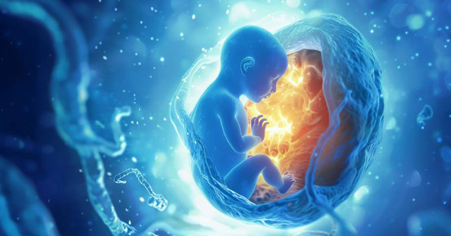
Recommendation
FISH provides exceptional clarity in detecting common aneuploidies while offering the significant advantage of speed - delivering answers within 24-48 hours when time is critical.
Our Excellence
At Suraksha Genomics, our FISH analysis for aneuploidy detection represents the pinnacle of cytogenetic precision:
Testing Process

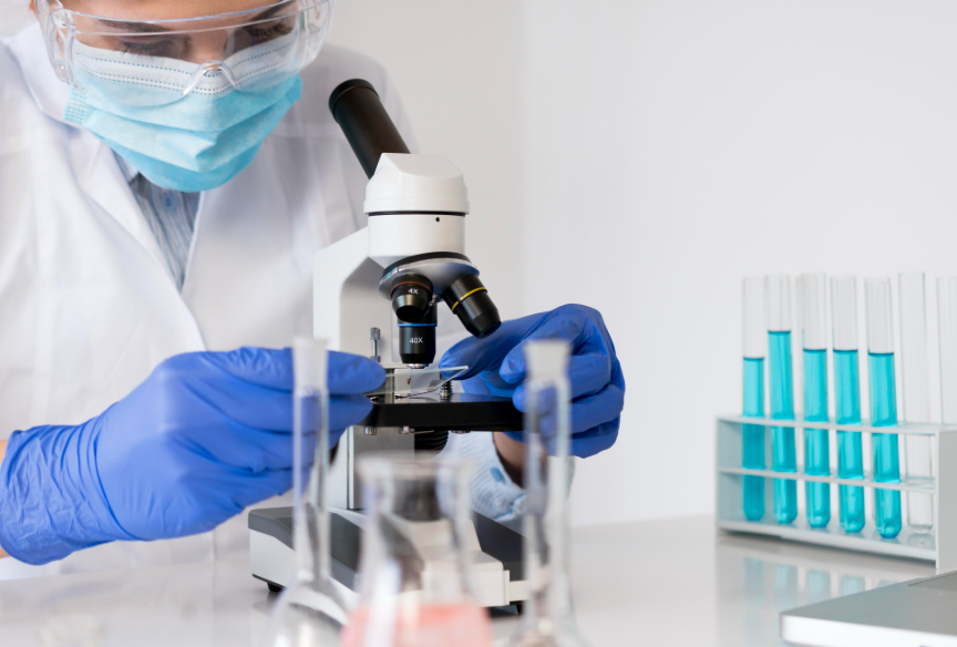
A small specimen is obtained by your healthcare provider through standard procedures.

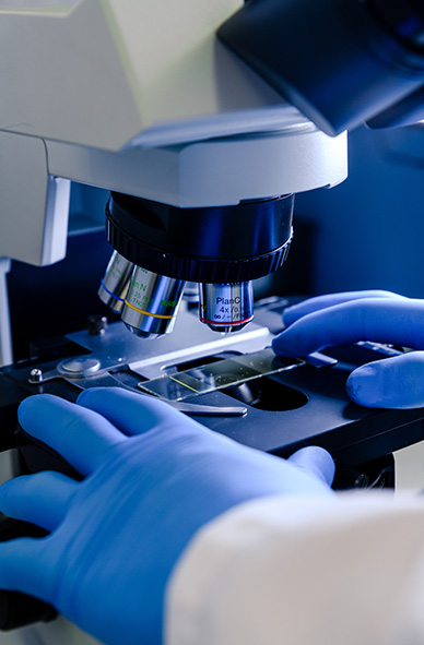
Our laboratory technicians isolate and fix cells on specialized slides.

DNA probes labeled with distinct fluorescent colors bind to specific chromosomes.

Specialized fluorescence microscopy reveals the number of each target chromosome.

Analysis of multiple cells by our cytogenetics specialists.

Clear, clinically relevant results delivered within 24-48 hour.

Expert consultation to explain findings and implications.
Why Suraksha Genomics?
your Result
FAQs
Fluorescence In Situ Hybridization (FISH) is a rapid genetic test used to quickly detect common numerical chromosomal abnormalities. It uses fluorescent probes that bind to specific chromosomes.
FISH is often used when rapid results are needed for suspected common aneuploidies, especially in prenatal samples (Amniotic Fluid or CVS) or in newborns with features suggestive of these conditions.
Our team understands the significance of genetic testing results and the impact they have on healthcare decisions. Our genetic counselors and laboratory specialists are available to answer your questions and provide supportive guidance throughout the testing process.
Contact our team today. Experience the clarity and confidence that comes from visual chromosome analysis by Eastern India's premier cytogenetics laboratory.
Consult our experts